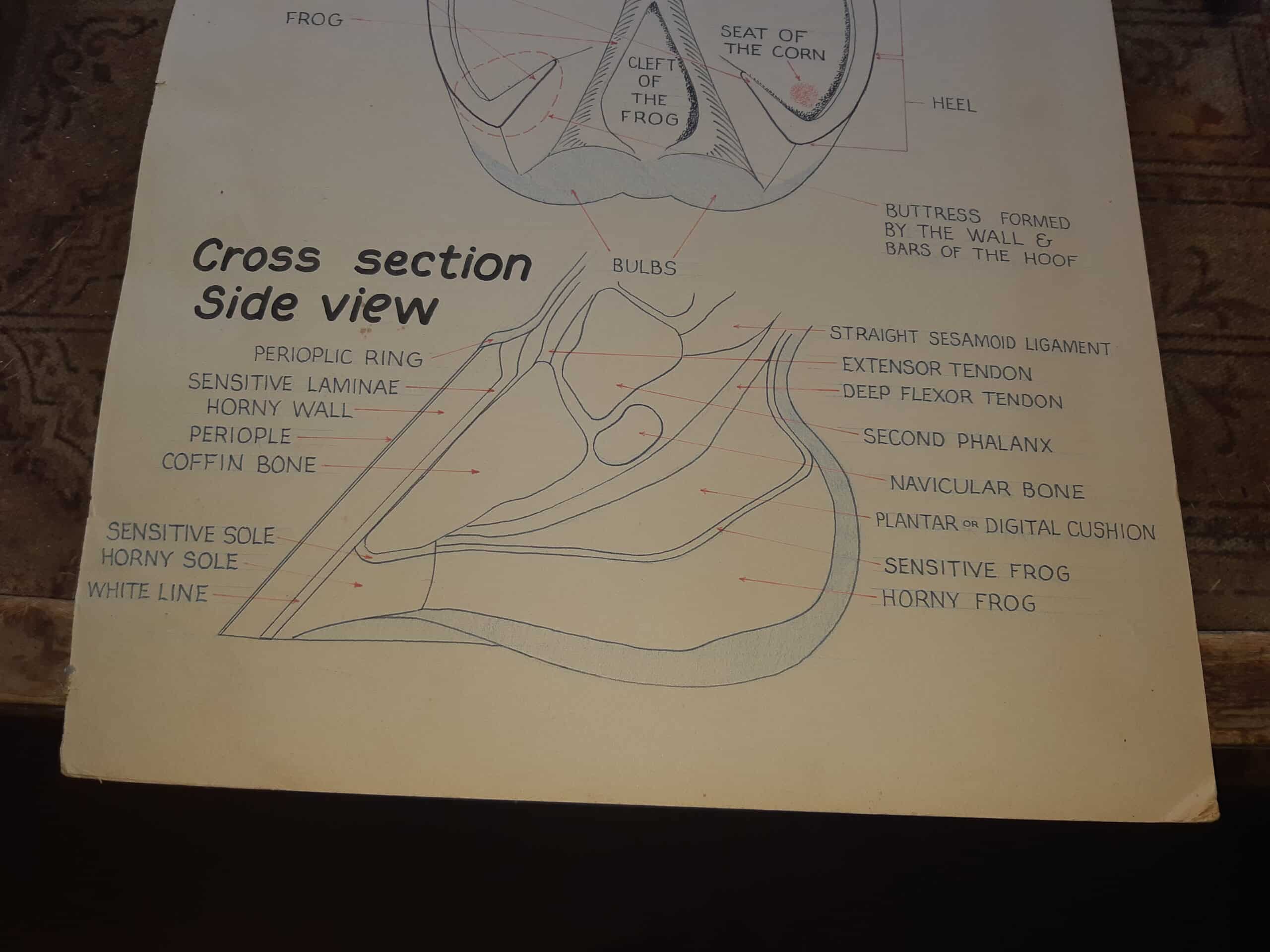
Navicular syndrome is a term used for chronic pain involving the structures at the back of a horse’s foot. It is more accurate to think of it as a syndrome than a single worn bone: the navicular bone, navicular bursa, deep digital flexor tendon, supporting ligaments and nearby soft tissues can all be involved.
Signs owners may notice
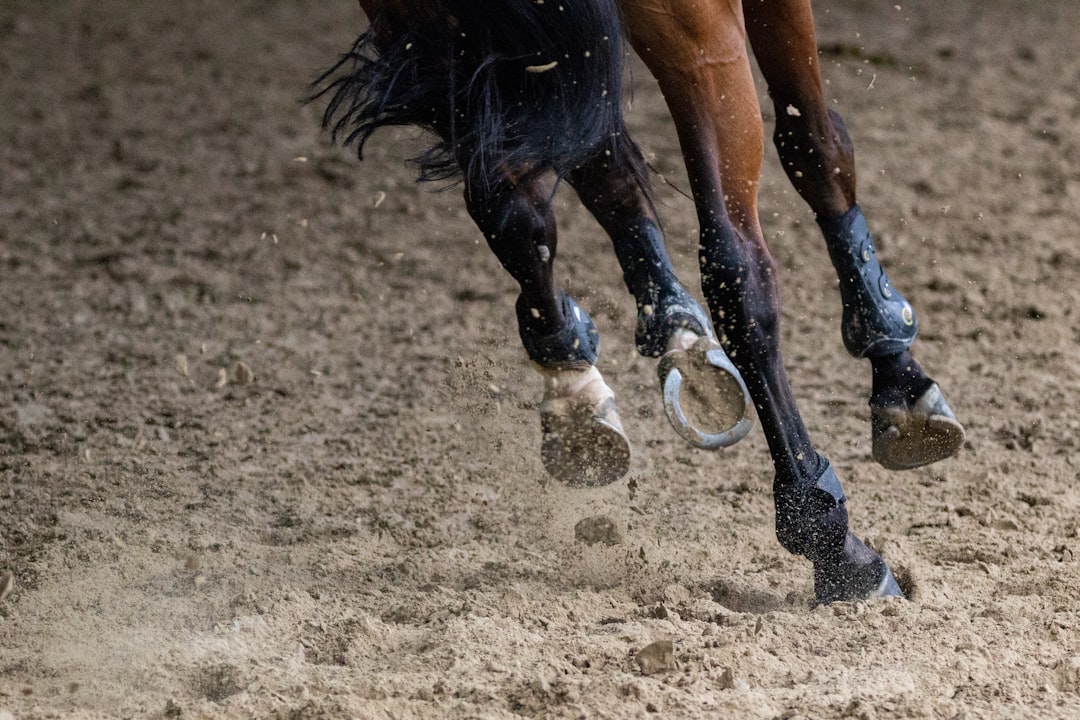
- A shortened or choppy stride, especially in front
- Intermittent lameness that appears to switch feet
- Stumbling, reluctance to extend the stride, or reduced performance
- Greater discomfort on a small circle, hard ground or after increased work
- Landing toe-first instead of loading the heel comfortably
These signs are not specific to navicular syndrome. Heel pain, sole bruising, hoof imbalance, arthritis, tendon or ligament injury and other conditions can look similar. A horse should not be labelled “navicular” from hoof shape or movement alone.
How navicular syndrome is diagnosed
A veterinarian begins with the horse’s history, a physical and lameness examination, hoof testers and observation on different surfaces. Diagnostic nerve blocks help localize pain to the foot. Radiographs can show changes in the navicular bone and hoof balance, but the severity of an X-ray change does not always match the horse’s pain.
When radiographs do not explain the lameness, ultrasound, MRI, CT or other advanced imaging may be considered. MRI is particularly useful for soft-tissue injuries inside the hoof capsule that ordinary X-rays cannot show.
Management is individualized
There is no single shoe, trim or medication that suits every case. Management may combine controlled exercise or rest, veterinary pain management, treatment of a specific soft-tissue injury, suitable footing, weight management and a farriery plan that reduces stress on painful structures.
Farriery commonly focuses on a comfortable, well-balanced foot, appropriate heel support and an easier breakover. The exact approach depends on the horse’s conformation, imaging, sole depth, current shoes, workload and response. Changes should be made deliberately and reassessed rather than assuming that more heel height or one therapeutic shoe will solve every case.
What owners can monitor
- Stride length and willingness to turn in both directions
- Whether the horse lands heel-first, flat or toe-first
- Comfort on soft versus firm ground
- Changes after shoeing, exercise, rest or medication
- Shoe wear, lost shoes and the interval at which balance begins to deteriorate
Practical farrier note: Good communication between the veterinarian, farrier and owner matters more than chasing a fashionable shoeing package. Radiographs taken with useful hoof markers can help the veterinarian and farrier plan the foot together.
Arrange a veterinary examination for persistent or recurring lameness. Masking pain and continuing full work can allow some foot injuries to worsen.
Related reading: Hoof balance and shoeing · Hoof bruising · Hoof anatomy
Veterinary reference: Merck Veterinary Manual: Navicular Syndrome in Horses.
Reviewed by Josh Emsley
Second-generation farrier serving Durham and Midwestern Ontario. Reviewed July 2026. This page provides general hoof-care information and does not replace an in-person veterinary diagnosis. View farrier services or contact Josh.