Horse Hoof Anatomy Made Simple
A practical guide for horse owners who want to understand what they are looking at without getting buried in veterinary terminology.
A horse’s hoof is more than a hard outer shell. It is a living, weight-bearing structure made of horn, soft tissue, blood vessels, nerves, tendons, and bone. You do not need to memorize every anatomical term to care for your horse well. The useful goal is to recognize the main parts, understand what they do and notice when something changes.
The hoof at a glance
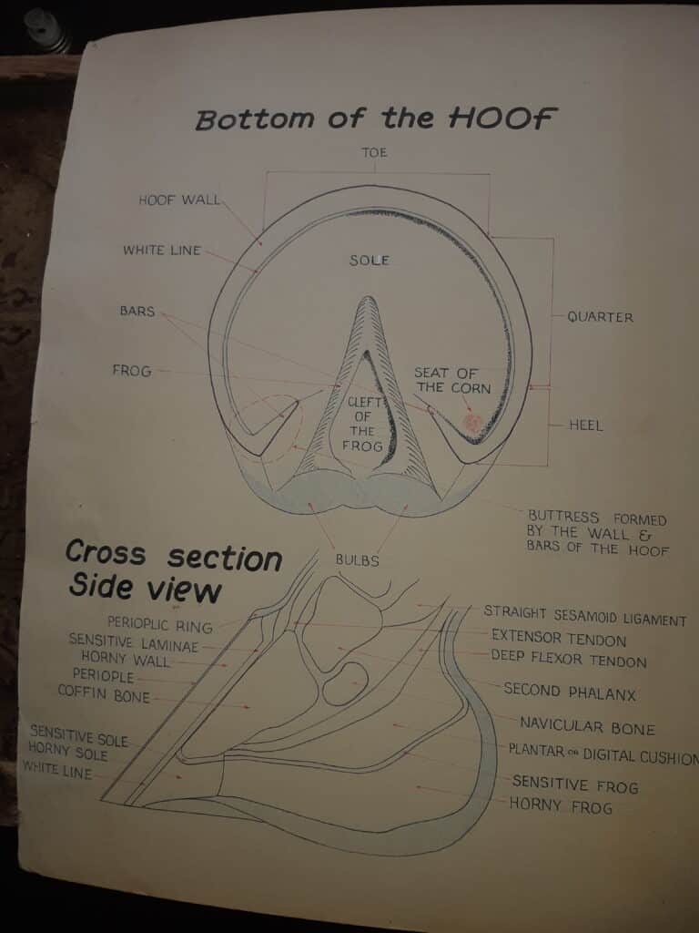
- Hoof wall: the hard outer part seen from the side.
- Sole: the protective underside of the foot.
- Frog: the V-shaped, rubbery structure in the centre.
- White line: the junction where wall and sole meet.
- Bars: inward folds of hoof wall beside the frog.
- Coronary band: the soft rim at the top where new wall grows.
- Inside the hoof: bone, laminae, joints, tendons and shock-absorbing tissues.
The parts you can see
Hoof wall
The hoof wall is the hard outer shell surrounding the foot. It protects sensitive structures and carries a large share of the horse’s weight. It grows downward from the coronary band, much like a fingernail grows from its nail bed.
Watch for: cracks, flares, uneven growth rings, chips, heat, tenderness or one side wearing differently.
Sole
The sole is protective horn covering most of the underside of the foot. A healthy sole is usually slightly concave rather than flat. Depending on the horse, footing and trim, parts of the sole may share load with the wall and frog.
Watch for: bruising, unusual softness, strong odour, punctures, packed stones or a flat, thin-looking sole.
Frog
The frog is the V-shaped, slightly rubbery structure in the centre of the foot. It contributes to traction, shock absorption and circulation within the hoof. A healthy frog should be firm but not rock-hard, with no deep painful split or foul-smelling black discharge.
Watch for: deep central cracks, tenderness, ragged loose tissue, black discharge, strong odour or a frog that is shrinking and no longer sharing load.
White line
The white line is the pale junction where the hoof wall and sole meet. Despite the name, it often looks cream or yellow. Stretching or separation here can allow dirt and infection to work upward into the wall.
Watch for: widening, crumbly horn, dark material, hollow areas, tenderness or separation near the toe or quarters.
Bars and heel area
The bars are inward folds of the hoof wall running beside the frog. Together with the heels and frog, they help support the back of the foot. Overgrown bars, contracted heels or a deep narrow central groove can contribute to discomfort and thrush.
Coronary band
The coronary band is the soft rim at the top of the hoof where new wall begins. Damage here can affect future hoof growth, so cuts, swelling or drainage near the coronet deserve prompt attention.
What is inside the hoof?
You cannot see these structures during routine hoof picking, but they explain why hoof care affects the whole limb.
- Coffin bone: the main bone inside the hoof capsule. It gives the foot shape and support.
- Laminae: thousands of tiny interlocking tissues that suspend the coffin bone inside the hoof wall. Laminitis damages this attachment.
- Digital cushion: a shock-absorbing pad beneath the back of the foot.
- Navicular area: a small bone and related soft tissues behind the coffin joint that help the deep digital flexor tendon move smoothly.
- Deep digital flexor tendon: runs down the back of the limb and attaches beneath the coffin bone.
- Blood vessels and nerves: nourish the living tissues and make deeper injuries painful.
Radiographs allow your veterinarian and farrier to compare the outside hoof capsule with the bones and sole depth inside. They are especially useful in laminitis, club feet, chronic lameness, severe cracks and difficult balance problems.
A 60-second owner hoof check
- Watch the horse walk before lifting the foot. Look for short steps, toe-first landings or reluctance to turn.
- Run your hand down the limb and compare heat or swelling with the opposite leg.
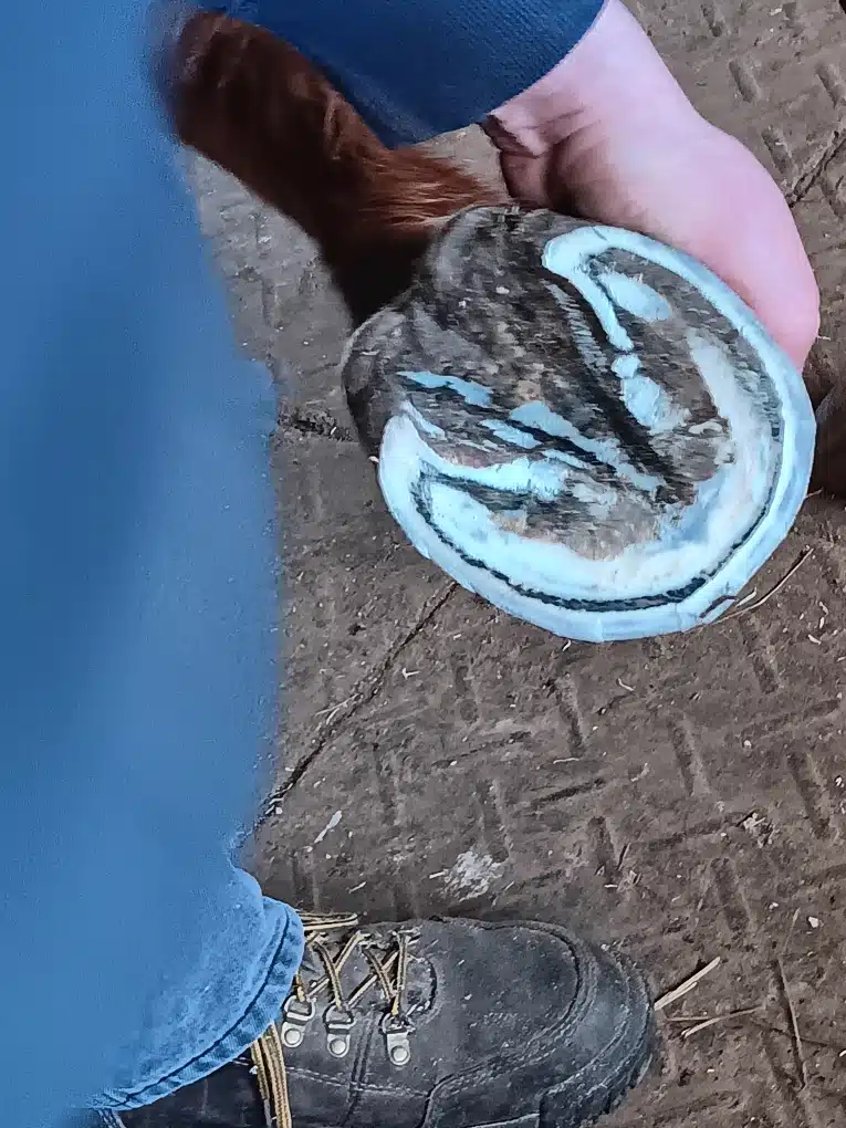
- Pick out the sole, frog grooves and heel area gently.
- Check for stones, nails, punctures, odour, discharge, bruising and loose wall.
- Compare all four feet. Sudden differences matter more than perfectly matching shapes.
- Notice whether shoes are tight, shifted, sprung or missing nails.
Taking occasional clear photographs from the front, side and sole can help you and your farrier track changes over time.
Call your farrier when
- The hoof is becoming long, flared or uneven between visits.
- A crack is widening, moving or reaching the coronary band.
- A shoe is loose, shifted, twisted or partly missing.
- The white line is stretching or crumbling.
- Thrush keeps returning despite cleaning and dry footing.
- You notice a steady change in hoof shape, landing pattern or comfort.
Call your veterinarian promptly when
- The horse is suddenly lame or will not bear weight.
- The hoof or lower limb is hot, swollen or strongly pulsing.
- There is a puncture, heavy bleeding or exposed sensitive tissue.
- Drainage, swelling or injury involves the coronary band.
- Laminitis is suspected, especially with a rocked-back stance or multiple painful feet.
- A problem is worsening despite appropriate farrier care.
The most useful takeaway
You do not need to identify every internal structure to be a good horse owner. Know the wall, sole, frog, white line, bars, heels and coronary band. Pick the feet regularly, compare them, and pay attention to changes in shape, smell, heat, comfort and movement. Early observations give your farrier and veterinarian a much better chance to solve small problems before they become major ones.
Related reading: Basic hoof care · How to pick out hooves · Common hoof problems
Reviewed by Josh Emsley
Second-generation farrier with more than 30 years of hands-on experience, serving Durham and Midwestern Ontario. Reviewed July 2026. This page provides general education and does not replace an in-person veterinary diagnosis. View farrier services or contact Josh.